-
-
-
-
-
-
Test for Fibroid
PELVIC EXAM
Uterine fibroids are often diagnosed by a pelvic exam. During this examination, fibroids may be determined by assessing the shape and size of the uterus. Further imaging studies, like ultrasound, may be utilized to confirm this finding.
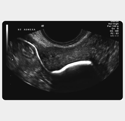
ULTRASOUND
Ultrasound uses the echoes from high-frequency sound waves to create a picture of the pelvic organs. As fibroids vary in size and location, both transvaginal and transabdominal ultrasounds may be used to best see the fibroids.3 D ultra sound is very useful in locating the exact site of the fibroid and its impact on the cavity. Doppler studies show the vascularisation of the fibroid. Adenomyosis is often difficult to distinguish from a fibroid. MRI – Magnetic resonance imaging is radiation-free imaging which gives a clearer picture of the uterus. The fibroids and the cavity of the uterus are better outlined.
SONOHYSTEROGRAPHY
Sonohysterography is an ultrasound procedure in which the uterine cavity is outlined by a small amount of fluid which is placed in the uterus through a thin plastic tube. Sonohysterography improves the doctor’s ability to identify fibroids which protrude into or distort the uterine cavity. The contrast echovist gives a clear outline of the cavity and the tubes.
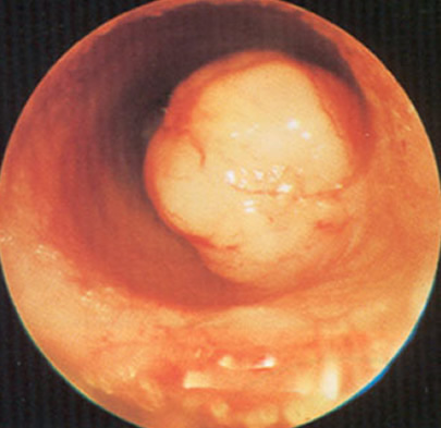
HYSTERO LAPAROSCOPY
HYSTERO LAPAROSCOPY involves direct examination of the uterus and its cavity (see endoscopy)

-

